
When you dream, your brain activates regions like the visual cortex, limbic system, and default mode network, which create vivid imagery, emotional content, and narrative flow. Meanwhile, areas responsible for logical judgment, like the prefrontal cortex, are less active, allowing surreal and spontaneous scenarios. During REM sleep, brain connectivity shifts significantly, supporting complex dreams and vivid memories. Continuing to investigate reveals more about how these neural patterns shape the dreams you experience.
How Do Dreams Form in the Brain During Sleep?
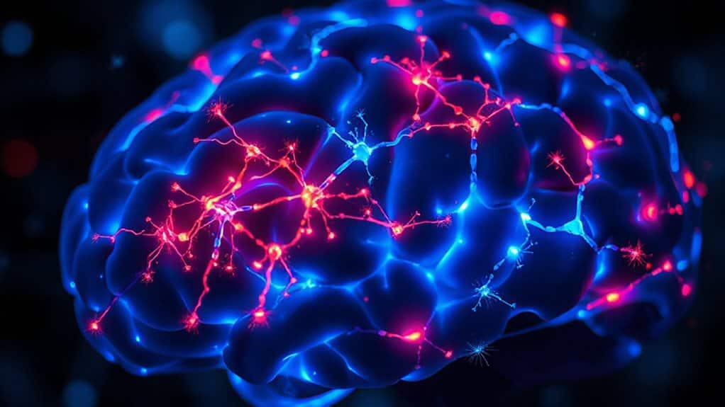
Dreams form in the brain through a complex interplay of neural activity that varies considerably during sleep, especially in REM sleep. During this stage, specific neural patterns activate the visual cortex, limbic system, and sensory areas, creating vivid imagery and emotions.
Brain activity in these regions supports dream formation while the prefrontal cortex remains less active, reducing logical judgment.
Which Brain Regions Are Active When You Dream?
When you’re dreaming, specific regions of your brain become highly active, shaping vivid experiences. The visual cortex generates detailed imagery, while the limbic system, including the amygdala, processes emotions.
The default mode network supports narrative and self-referential thought, whereas the prefrontal cortex, responsible for judgment, remains less active, contributing to the dream’s often illogical nature. Dreams may also reflect subconscious cues about tension, anxiety, or emerging issues.
How Do Sleep Stages Influence Dreaming?
Different sleep stages considerably influence the nature and intensity of dreaming. During REM sleep, brain activity increases, especially in the visual cortex and limbic system, leading to vivid imagery and emotional dream content.
Non-REM stages involve slower brain waves and less intense dreaming, resulting in more static dreams.
Longer REM episodes later in the night produce more elaborate and memorable dreams. Sleep paralysis can occur when REM atonia briefly carries into wakefulness, leaving you mentally aware but unable to move.
How Do Brain Networks Create Dream Content and Visual Imagery?
During dreaming, your brain’s networks activate in specific patterns that produce vivid visual imagery and complex narratives. The visual cortex, especially in the occipito-temporal region, becomes highly engaged, creating the images you see in your dreams.
Meanwhile, the default mode network helps construct the storylines and self-referential thoughts. Additionally, activity in regions like the temporo-parietal junction can influence your level of self-awareness, enabling phenomena like lucid dreaming or some degree of control over dream content.
Neural Activation Patterns
The creation of dream content and visual imagery results from complex patterns of neural activation across multiple brain networks. During dreaming, neural activation in the visual cortex increases, supporting vivid imagery.
Brain networks like the limbic system heighten emotional and memory-related activity, while the prefrontal cortex deactivates, allowing surreal scenarios to unfold without logical constraints.
Visual Cortex Engagement
When you dream, your brain’s visual cortex becomes highly active, playing a key role in generating the vivid imagery experienced during REM sleep.
Neural activity in this region creates detailed scenes, faces, and objects by processing complex shapes, colors, and spatial relationships.
This heightened activity occurs despite reduced frontal lobe activity, allowing for immersive, surreal visual content driven by neural networks.
Self-Awareness and Control
How do brain networks generate the vivid and often surreal content of dreams, especially in the absence of conscious control? During REM sleep, the prefrontal cortex, responsible for self-awareness and volition, is less active, reducing logical thinking.
In lucid dreaming, increased activity in the temporo-parietal junction allows for self-awareness and conscious influence over dream content.
Why Is REM Sleep Necessary for Vivid Dreams?
REM sleep is essential for producing vivid dreams because it activates specific brain regions responsible for sensory and emotional processing. During REM, the visual cortex and limbic system become highly active, supporting detailed imagery and emotional intensity.
This synchronization of brain activity enables the surreal and emotionally charged qualities of vivid dreams, which are less likely outside of REM sleep.
How Does the Limbic System Shape Dream Emotions?
The limbic system plays a central role in shaping the emotional terrain of your dreams, especially during REM sleep when it becomes highly active.
The amygdala, a key part of this system, processes intense emotional experiences like fear, joy, or anger. Its heightened activity results in vivid dream emotions, often without the critical judgment of the prefrontal cortex.
Because dreams about a damaged car often reflect feelings of loss of control, the emotions you wake with can offer clues about what your brain is processing.
How Does Brain Activity Differ Between Wakefulness and Dreaming?
Although sleep might seem like a passive state, your brain remains remarkably active, especially during dreaming. During REM sleep, brain activity increases in visual, limbic, and associative cortices, resembling wakefulness.
Cortical activation is uneven, with reduced activity in the prefrontal cortex, which explains the illogical nature of dreams. The limbic system’s heightened activity influences emotions and memory processing.
Can Brain Lesions Predict Changes in Dreaming Experience?
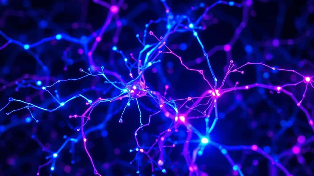
Research indicates that brain lesions can markedly influence dreaming experiences by disrupting specific neural regions involved in visual imagery, self-awareness, and emotional processing.
Damage near the temporo-parieto-occipital junction impairs mental imagery and visuo-spatial abilities, while lesions in the limbic system alter emotional content.
These injuries help predict changes in dreaming, linking affected brain areas to specific dream features.
For some people, a calming amethyst crystal placed near the bed is also used in nighttime rituals to support relaxation and dream recall.
What Do Neuroimaging Studies Reveal About Dream Generation?
Neuroimaging studies reveal that during dreaming, specific brain regions become highly active, especially those involved in visual processing, emotion, and memory, such as the occipito-temporal cortex, amygdala, and hippocampus.
Meanwhile, areas responsible for logical reasoning and self-awareness, like the prefrontal cortex, are particularly deactivated, contributing to the dream’s often illogical nature.
These studies also highlight complex connectivity patterns across brain networks, suggesting that dream generation involves widespread, fluid neural interactions similar to wakefulness, but with distinct regional modulations.
Some researchers also note that the altered awareness seen in lucid dreaming can help illuminate how the brain shifts between passive dream creation and conscious control.
Brain Activation Patterns
Have you ever wondered what happens in your brain when you dream? Neuroimaging studies show that during REM sleep, brain activation occurs mainly in emotion, memory, and visual areas.
Meanwhile, the prefrontal cortex is markedly deactivated. This pattern explains the vivid visuals and surreal quality of dreams, as logical reasoning and self-awareness are temporarily suppressed.
Key Dream Regions
What brain regions are most involved in generating dreams? Neuroimaging studies reveal that during REM sleep, the limbic system, including the amygdala and hippocampus, becomes highly active, supporting emotional and memory processing.
The visual cortex is also engaged, creating vivid imagery.
Meanwhile, the prefrontal cortex remains subdued, reducing logical reasoning and self-awareness, which contributes to the surreal quality of dreams.
Brain Connectivity Dynamics
During REM sleep, the brain operates as a lively and highly interconnected network, with shifts in regional connectivity shaping dreams. Neuroimaging studies, especially functional MRI, reveal increased connectivity between regions like the hippocampus and visual cortex, supporting vivid imagery and memory integration.
Meanwhile, deactivation of the prefrontal cortex results in diminished logical reasoning, highlighting flexible brain connectivity during dreaming.
Wrapping Up
Certainly! Here’s the modified text with alternative words replacing “in summary,” “insights,” and “insight”:
In brief, understanding how your brain generates dreams involves examining specific regions, sleep stages, and neural networks. Brain activity during REM sleep, especially in the limbic system, contributes to the vividness and emotional content of dreams. Neuroimaging and studies of brain lesions offer perspectives into these processes, revealing that dreaming is a complex interplay of multiple brain functions. This knowledge deepens our comprehension of sleep’s role in mental processes, though many aspects remain an intriguing frontier for future research.

Seraphina Lumina is a dream interpretation and symbolism researcher with a passion for exploring the connections between dreams, emotions, relationships, spirituality, and personal growth. She draws on traditional dream symbolism, psychological perspectives, and spiritual traditions to help readers make sense of their night-time experiences and the themes that may appear in them.